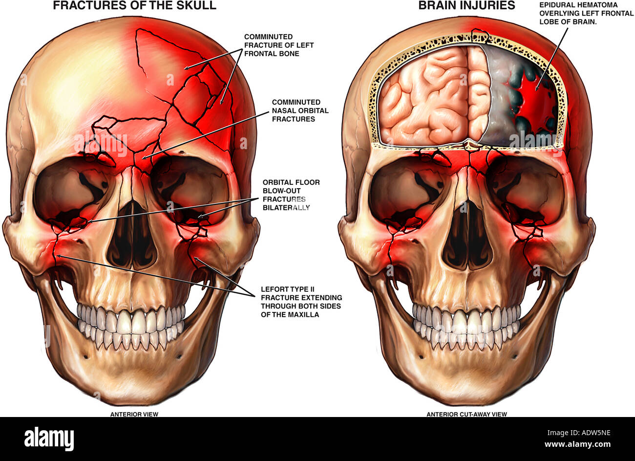


It is particularly important to document facial nerve function with penetrating trauma to the lateral face.Īssess for anesthesia due to injury of various branches of the trigeminal nerve by lightly touching the forehead, lower eyelid, cheek, upper lip, and chin. (5,14)Įvaluate facial motor function by having the patient close their eyes tightly, raise their eyebrows, purse their lips, smile, and frown. Periorbital ecchymoses, or raccoon eyes, are associated with various injuries including basilar skull, nasoorbitoethmoid, Le Fort, orbital, and frontal bone fractures. Perform a thorough inspection of the face and oropharyngeal cavity with both a “bird’s eye view” from above and a “worm’s eye view” from below to evaluate for subtle facial asymmetries.

How is your vision (blurriness, double vision, floaters or flashes of light, photophobia, foreign body sensation, pain with eye movements, etc.)?ĭo your teeth fit together normally? (14)Īll patients should also be asked about the use of anticoagulants or platelet-inhibiting medications. (13,15,16)įocused History - The following three screening questions can be used to help localize injuries: (1,15) Life-threatening hemorrhage not controlled by these interventions may require emergent arterial ligation or embolization. (1,14,15) In particular, epistaxis in midfacial fractures can be controlled with posterior nasal packing with nasal tampons, Foley catheters, double lumen balloon catheters, or other nasal packing materials. (1) Control hemorrhage early with direct pressure and early packing of the nasal and oral cavity as needed. (5,11-13) Severe hemorrhage occurs most commonly in midfacial fractures with the maxillary artery and its intraosseous branches as the origin of bleeding. (5) Severe and life-threatening hemorrhage in maxillofacial trauma ranges has a reported incidence up to 11%. Massive hemorrhage, significant enough to precipitate shock, can occur with maxillofacial trauma due to increased vascularity of facial structures. At minimum, cervical spinal precautions should be maintained as the incidence of cervical spinal injury associated with facial fractures ranges from 0.3 to 24.0%. (8,9) However, this is not always feasible as patients often must maintain spinal precautions during the initial stages of their evaluation. Allowing patients to sit up can allow them to maintain their own airway and prevent aspiration of blood and emesis. Lastly, it is important to consider optimal positioning of patients requiring airway intervention. (6) If bag-mask ventilation becomes difficult, consider using a supraglottic airway device as a bridge to a definitive airway.Īdditionally, given high occurrence of soiled airways in maxillofacial trauma, providers must ensure multiple suction catheters (preferably large-bore DuCanto catheters) are at the ready and should consider advanced intubation methods such as Suction Assisted Laryngoscopy and Airway Decontamination (SALAD, discussed in further detail in this post). When considering airway interventions, providers should anticipate difficulty in bag-mask ventilation as distorted facial anatomy can prevent adequate mask seal (particularly in Le Fort II and III fractures). (4) Airway compromise most commonly occurs due to soiling of the airway (significant hemorrhage or emesis) and obstruction (posteriorly displaced tongue, soft tissue injury and swelling, or other foreign bodies such as dislodged teeth). In the setting of maxillofacial trauma, airway compromise and severe hemorrhage are the most common life-threatening complications.Įnsuring airway patency is of the utmost importance in patients with maxillofacial trauma as up to 42% of patients with severe maxillofacial trauma require intubation.


 0 kommentar(er)
